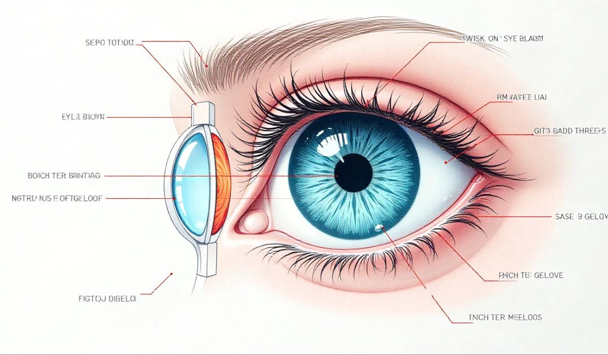
In the meticulous world of cataract surgery, one truth stands above all: the foundation of a perfect refractive outcome is precise biometry. An error of just a fraction of a millimeter in axial length can translate into a significant and disappointing postoperative refractive surprise for your patient. For decades, ophthalmic professionals have relied on A-scan ultrasound to provide this critical data, but traditional devices often came with a steep learning curve and inherent variability.
Enter the DGH A (Scanmate A), a next-generation, portable A-scan ultrasound biometer engineered to eliminate these common pitfalls. This guide will provide you with a full understanding of this device’s technical capabilities, practical usage modes, and profound clinical benefits in surgical planning and myopia management. You will see why the DGH A is rapidly becoming the standard for portable, precise ophthalmic measurements in high-volume clinics, surgery centers, and remote eye camps alike.
Understanding DGH A: The Foundation of Ophthalmic Biometry
Before delving into its advanced features, it’s crucial to grasp the core function and fundamental importance of the DGH A in clinical practice.
What is the DGH A (Scanmate A) and its Core Function?
The DGH A is a sophisticated A-scan ultrasound biometer that utilizes a high-frequency 10 MHz probe to make precise, time-amplitude measurements of the internal structures of the eye. Its primary role is to accurately measure the axial length measurement—the distance from the corneal surface to the retinal pigment epithelium—within a robust 15–40 mm range. Additionally, it provides critical data on lens thickness measurement, anterior chamber depth, and vitreous length.
These measurements are not merely numbers on a screen; they are the direct inputs for IOL power calculation. The accuracy of these readings directly dictates the power of the intraocular lens you will implant, making the DGH A one of the most influential tools in your surgical planning arsenal.
Why Accuracy Matters: Biometry for Refractive Outcomes
To underscore the device’s importance, consider the direct clinical impact of its measurements. An error of just 0.1 mm in axial length can lead to a 0.25–0.30 D miscalculation in IOL power. In an era where patients expect emmetropia, such a deviation is often unacceptable.
Beyond cataract surgery, the DGH A plays a vital role in myopia tracking. In pediatric ophthalmology, monitoring the elongation of the axial length over time provides an objective, critical measure of myopia progression. This allows you to gauge the effectiveness of control strategies (like atropine or specialty contact lenses) far more reliably than relying on refractive changes alone. The device’s reliability is backed by its FDA and CE approvals, providing a trusted foundation for these sensitive clinical decisions.
The Two Modes: Contact vs. Immersion A-Scan
The DGH A supports two primary measurement techniques, each with distinct advantages:
- Contact Mode: This is a direct application method where the probe tip touches the cornea (under topical anesthesia). While fast and efficient, it carries an inherent risk of corneal compression, which can artificially shorten the axial length and lead to a hyperopic surprise postoperatively.
- Immersion Mode: Widely considered the gold standard for A-scan accuracy, this technique uses a saline-filled shell (like a Prager Shell) placed on the eye. The probe scans through the fluid medium without touching the cornea. This immersion biometry technique virtually eliminates corneal compression error, providing superior and more reliable data.
A common question we hear is, “What is the difference between contact and immersion A-scan biometry with the DGH A?” The answer lies in the compromise between speed and absolute accuracy. Contact is faster, but immersion is demonstrably more accurate and reproducible, making it the preferred method for achieving premium refractive targets.
Essential Features That Elevate the DGH A Workflow
The DGH A isn’t just an accurate machine; it’s an intelligently designed tool that enhances user efficiency and patient safety.
Safety and User Guidance Systems
One of the most significant advancements in the DGH A is its integrated guidance and safety features, which standardize performance across all levels of user experience.
- Star and Sound Guidance: This real-time feedback system is a game-changer for training and efficiency. The device’s display shows a star pattern and emits an auditory signal that changes in pitch. The user simply adjusts the probe position until the star is perfectly formed and the tone is at its highest pitch, indicating optimal alignment and signal quality. This removes the guesswork and significantly shortens the learning curve for new technicians.
- Safety Lock (Compression Lockout): This critical feature automatically prevents a measurement from being taken if the probe is applying excess pressure to the eye. This DGH A compression lockout feature protects the patient from discomfort and potential corneal injury while simultaneously safeguarding the integrity of your biometry data by preventing artificially shortened readings.
Portability and Integration
Weighing in at less than one pound, the DGH A handpiece is the epitome of portability. It connects via a standard USB 2.0 cable to any Windows-based PC or laptop, transforming any space into a fully functional biometry station.
This makes it the ideal portable A-scan ultrasound machine for eye camps, satellite offices, nursing homes, and operating rooms where a permanent, bulky unit is impractical. Its plug-and-play setup means you can be up and running in minutes, bringing high-quality eye care to underserved populations without compromising on data quality.
Technical Specifications for High Reliability
Underpinning its user-friendly design are robust technical specifications that ensure clinical trust:
- High Resolution and Consistency: The device offers a measurement resolution of 0.01 mm and boasts an consistency of ±0.03 mm, ensuring that your results are not only precise but also highly repeatable across multiple operators and visits.
- Plug-and-Play Simplicity: Unlike older, complex machines with proprietary hardware, the DGH A leverages modern computing, simplifying setup, maintenance, and software updates.
Advanced Clinical Applications and IOL Power
The true value of the DGH A is realized in its advanced software capabilities, which empower you to handle a wide range of clinical scenarios.
Accessing Comprehensive IOL Power Calculation Formulas
The integrated software is a powerhouse of clinical intelligence. It comes pre-loaded with all major industry-standard IOL calculation formulas, including SRK/T, Hoffer Q, Haigis, Holladay 1 and 2, and Shammas. This allows you to compare formulas and select the best one for each patient’s unique ocular dimensions.
Crucially, it also includes specific formulas and modules for calculating IOL power in post-refractive surgery eyes. This directly addresses the complex challenge of how the DGH A calculates IOL power for post-LASIK patients, helping to mitigate the risk of refractive surprises in this demanding population.
Utilizing DGH A for Pediatric Myopia Management
The application of the DGH A extends far beyond the operating room. Its software includes functionality to generate and track axial length growth charts over multiple patient visits. By plotting a child’s axial length against normative data, you can objectively monitor the rate of elongation. This is a cornerstone of modern myopia tracking, allowing for early intervention and data-driven assessment of treatment efficacy, making the DGH A an invaluable tool in pediatric ophthalmology.
When to Choose the DGH A Over Optical Biometers
While optical biometers (like the IOLMaster or Lenstar) are excellent tools, they have limitations in dense or opaque media. This is where the DGH A proves to be an indispensable asset.
What makes the DGH A the preferred portable A-scan device for mobile clinics? Its reliability in the face of challenging pathology. In cases of dense white cataracts, significant corneal edema, scarred corneas, or in patients with extreme fixation issues (including children and those with nystagmus), optical coherence-based systems often fail to acquire a signal. Ultrasound, as utilized by the DGH A, penetrates these opaque media with ease, ensuring you can obtain the vital axial length data needed to proceed safely with surgery. It is not just a backup; for complex eyes, it is often the primary solution.
Conclusion
The DGH A (Scanmate A) represents a significant evolution in ophthalmic biometry. It successfully merges the gold-standard accuracy of immersion ultrasound with unparalleled portability and intelligent user-assist features. By providing reliable, reproducible measurements of axial length and lens thickness, it empowers cataract surgeons to achieve superior refractive outcomes. Furthermore, its utility in pediatric myopia management and its reliability in challenging clinical situations make it a versatile and essential tool for any modern ophthalmic practice, regardless of size or location.
This device empowers your clinic with accurate data, leading to better patient outcomes and a more efficient workflow. To fully appreciate its capabilities, we encourage you to explore the DGH A firsthand. Contact us or your representative to request a live product demo or inquire about specialized training for your clinical staff to integrate this powerful tool into your practice seamlessly.
YOU MAY ALSO LIKE: Promotional Gifts: Smart Strategies to Make Your Brand More Visible

